

When people think about stroke, they often picture a sudden emergency. A person collapses, symptoms appear quickly, and immediate action is required. In those moments, imaging decisions are made under pressure, with speed as the top priority.
At the same time, stroke risk does not always announce itself dramatically. In many cases, underlying changes develop quietly, long before any obvious warning signs appear. This creates confusion around imaging. If CT is commonly used for stroke, why is MRI often discussed in preventive screening? And if MRI can reveal more detail, why isn’t it used in emergencies?
This article clarifies that CT and MRI serve different purposes in stroke care. Rather than asking which test is better, it explains how each tool fits a specific goal, whether that goal is urgent decision-making or proactive risk assessment.
Why CT and MRI Are Often Compared in Stroke Care
Both are brain imaging tools, but with different goals
CT and MRI are both widely used to image the brain. Because they can each be involved in stroke-related care, they are often grouped together in conversations about detection and screening.
This grouping can create the impression that CT and MRI are interchangeable, or that one is simply a more advanced version of the other. In reality, they are designed to answer different clinical questions. Understanding those questions is more useful than focusing on the machines themselves.
Confusion between emergency care and prevention
Stroke is commonly associated with emergency treatment. In that context, imaging is used to guide immediate decisions. However, stroke risk can exist long before an emergency occurs.
When discussions shift from acute events to prevention, the purpose of imaging changes. The goal is no longer to respond to a crisis, but to understand risk, patterns, or subtle changes that may influence long-term planning. Mixing these two contexts is one of the main reasons CT and MRI are often misunderstood when compared.
How CT Is Used in Suspected Stroke
Speed matters in emergency situations
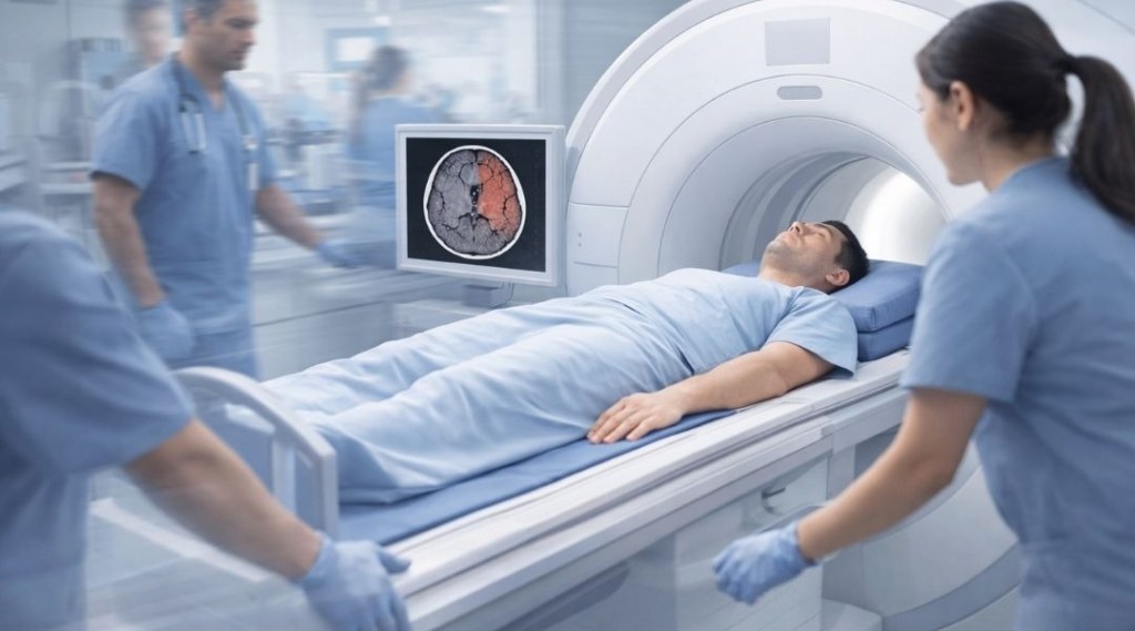
In emergency settings, time is critical. When a stroke is suspected, clinicians need information quickly to guide immediate decisions. CT is widely used in these situations because it can be performed rapidly and is readily available in most emergency departments.
The priority in this context is not long-term risk assessment, but fast clarification. Imaging needs to support urgent action, often within minutes, rather than provide detailed insight into subtle or long-standing changes.
Detecting acute bleeding and major changes
One of the key roles of CT in suspected stroke is identifying acute bleeding or large structural changes in the brain. This information helps clinicians distinguish between different types of stroke-related events and determine next steps in emergency care.
CT is not used here as a screening tool. Its value lies in answering a specific, time-sensitive question: is there an acute issue that requires immediate intervention? This purpose explains why CT remains central in emergency stroke workflows, even though other imaging tools may offer greater detail in different contexts.
How MRI Is Used in Stroke Screening and Prevention
Seeing subtle changes that develop quietly
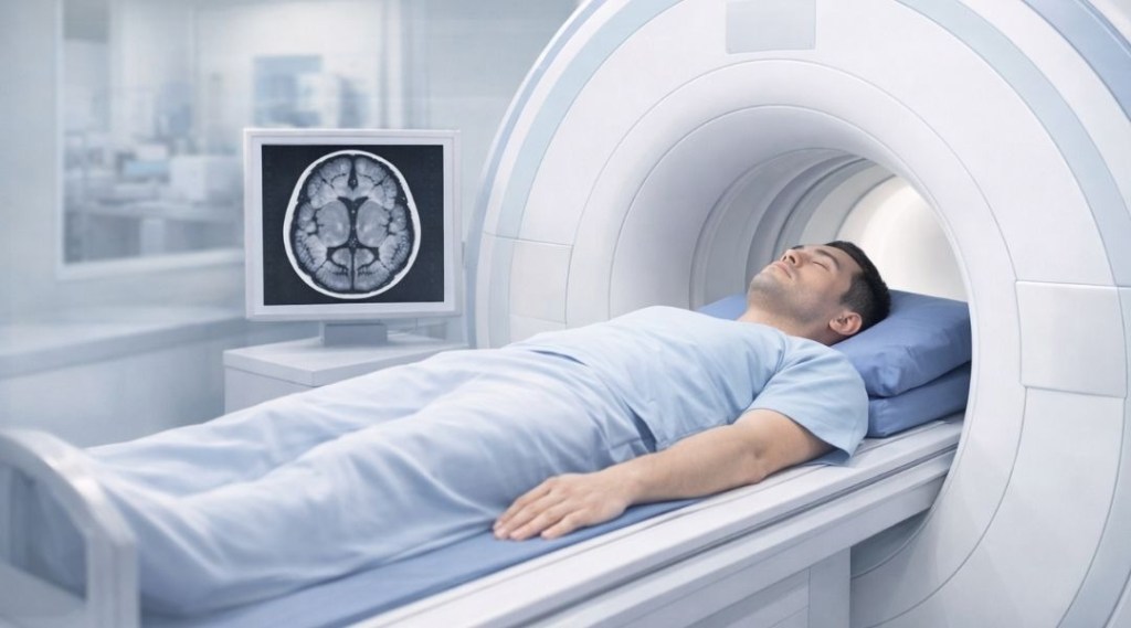
Unlike emergency imaging, preventive screening focuses on changes that may not cause immediate symptoms. MRI is often discussed in this context because it can reveal subtle structural changes in the brain that develop gradually over time.
These may include small areas of tissue change or patterns that are less visible on faster imaging methods. The emphasis is not on confirming a stroke event, but on gaining visibility into changes that could be relevant for monitoring or further evaluation.
This ability to detect finer detail is why MRI is frequently mentioned in conversations about stroke risk, even when no acute symptoms are present.
MRI in proactive stroke risk assessment
When the goal is proactive assessment rather than emergency response, imaging serves a different purpose. MRI may be considered as part of broader efforts to understand risk factors or establish a baseline for future comparison.
This does not mean MRI is automatically used for screening, or that it is appropriate for everyone. Its role depends on context, including medical history and the specific question being explored. In preventive settings, MRI contributes information that supports planning, rather than triggering immediate action.
Emergency Imaging vs Preventive Screening
Different questions require different tools
The key difference between CT and MRI in stroke care is not the technology itself, but the question being asked.
In an emergency, imaging is used to determine whether immediate action is needed. The priority is speed and clarity in a high-pressure situation. CT is well suited to this role because it can quickly identify major findings that influence urgent decisions.
In preventive screening, the question is different. Instead of asking whether a stroke is happening right now, the focus is on whether there are signs that warrant monitoring or further evaluation. MRI aligns more closely with this goal because it provides detailed insight into subtle changes that may develop quietly over time.
Why one does not replace the other
Because CT and MRI answer different questions, one does not replace the other.
Using CT for prevention would miss the purpose of emergency imaging, just as relying on MRI in an acute emergency would not match the need for speed. Each tool is effective within its intended context, and confusion arises when those contexts are blended together.
Understanding this separation helps explain why both CT and MRI remain essential in stroke care, even though they are used at different stages and for different objectives.
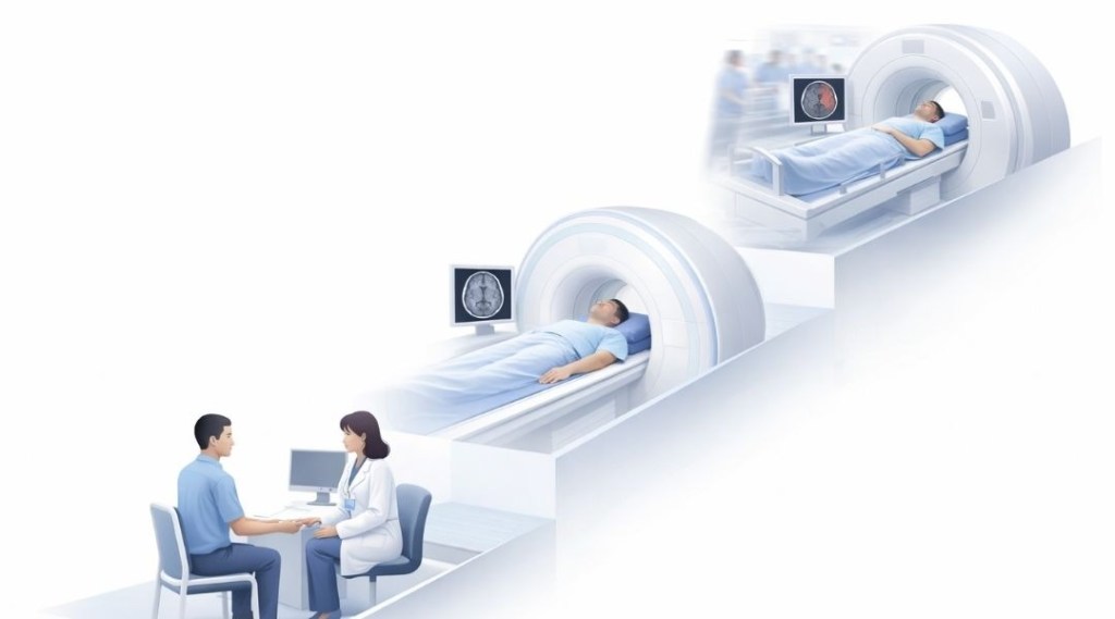
What “Stroke Screening” Actually Means
Screening focuses on risk, not diagnosis
In this context, the word “screening” can be misleading if it is not clearly defined.
Stroke screening does not mean confirming that a stroke has occurred, and it does not provide a diagnosis. Instead, screening looks for indicators that may suggest increased risk or changes that are worth observing over time. The intent is to support awareness and planning, not to label a condition.
This distinction matters because imaging used for screening is interpreted differently from imaging used in emergencies. The results are considered alongside other information, rather than treated as definitive answers.
Why screening is not the same as emergency imaging
Emergency imaging is driven by urgency. It is designed to answer immediate questions that affect rapid decision-making.
Screening, by contrast, is slower and more contextual. Timing, intent, and follow-up expectations are different. Imaging in this setting aims to provide visibility, not resolution. Mixing these two purposes can lead to confusion about what a scan can realistically offer.
Recognizing this difference helps set appropriate expectations and reduces the risk of misinterpreting screening results as emergency findings.
Choosing Imaging Based on the Goal
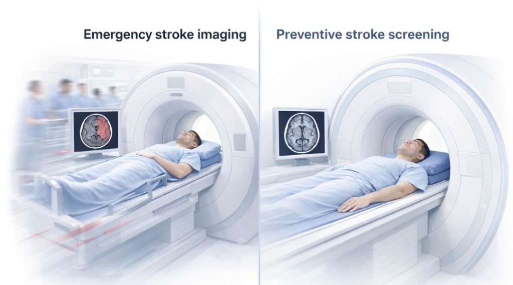
When urgency is the priority
When stroke is suspected as an active emergency, the primary goal is to make time-sensitive decisions. Imaging in this situation supports rapid assessment rather than long-term understanding.
In these moments, the question is whether immediate intervention is needed. Speed, availability, and clarity take precedence. This context explains why CT remains closely associated with emergency stroke care and why it is not used as a general screening tool.
When prevention and planning are the focus
When the goal shifts from urgency to prevention, the role of imaging changes.
In proactive discussions, imaging may be used to understand potential risk, establish a baseline, or explore subtle changes that could inform future monitoring. MRI is often discussed in this setting because its strengths align with detailed evaluation rather than rapid triage.
This difference in purpose reinforces an important point: imaging choices are driven by intent, not by a hierarchy of tests.
How to Think About CT and MRI Together
Complementary roles in stroke care
CT and MRI are best understood as complementary tools rather than competing options.
CT plays a critical role when rapid answers are needed. MRI contributes when deeper insight and contextual understanding are the priority. Each supports a different stage of care, and each is used most effectively when applied to the question it is designed to answer.
Seeing them together in this way helps reduce the tendency to frame one as superior to the other.
Imaging as part of a structured approach
Imaging does not exist in isolation.
CT and MRI are used within broader frameworks that include clinical evaluation, history, and follow-up planning. When discussed in preventive contexts, MRI fits within a structured preventive stroke screening approach, where tools are used sequentially and thoughtfully rather than as standalone solutions.
This perspective keeps expectations grounded and emphasizes the role of context in interpreting imaging results.
Key Takeaway
CT and MRI serve different purposes in stroke care because they answer different questions. CT supports fast decision-making in emergencies, while MRI offers detailed insight that can be useful in preventive screening and risk assessment. Understanding this distinction helps clarify why imaging choices depend on goals rather than on finding a single “best” test.
FAQs – CT vs MRI for Stroke Screening
1. Is CT or MRI better for stroke screening?
There is no single “better” option.
CT and MRI serve different purposes. CT is primarily used in emergency situations, while MRI is often discussed in preventive or screening contexts where the goal is understanding risk rather than responding to an acute event.
2. Why is CT commonly used first in suspected stroke emergencies?
CT is widely used in emergencies because it is fast and accessible.
In urgent situations, imaging needs to support immediate decision-making, especially when there is concern about acute bleeding or major structural changes.
3. Can MRI detect stroke risk before symptoms appear?
MRI may reveal subtle brain changes that develop quietly over time.
In preventive discussions, this visibility can support risk assessment or monitoring, but it does not diagnose a stroke or predict when one might occur.
4. Does CT work for preventive stroke screening?
CT is not typically used as a preventive screening tool.
Its primary role is to clarify urgent findings in acute settings, not to assess long-term stroke risk when symptoms are absent.
5. Why isn’t MRI used in emergency stroke situations?
MRI usually takes longer and is less suited to rapid decision-making in emergencies.
When time is critical, imaging methods that can be performed quickly are prioritized over those designed for detailed evaluation.
6. What does “stroke screening” actually aim to do?
Stroke screening focuses on identifying indicators that may suggest increased risk.
It does not confirm a stroke diagnosis and does not replace emergency imaging or clinical evaluation.
7. Can CT and MRI be used together in stroke care?
Yes, but at different stages and for different goals.
CT and MRI are complementary tools. Each contributes information that is relevant to a specific context rather than serving the same function.
8. If I have risk factors, should I choose MRI instead of CT?
Imaging choices depend on context and medical guidance.
Risk factors may influence discussions about preventive screening, but imaging decisions are not made based on risk alone and should always consider the clinical situation.
9. Does a normal MRI or CT mean I will not have a stroke?
No.
Imaging provides information at a specific point in time. It does not offer certainty about future events and must be interpreted alongside other factors.
10. What is the key takeaway when comparing CT and MRI for stroke screening?
CT and MRI answer different questions.
CT supports rapid decisions in emergencies, while MRI can provide deeper insight in preventive contexts. The appropriate choice depends on the goal, not on which test is considered more advanced.
VIETWELLNESS TOUR
Plan Your Full-Body MRI
Doctor-led preventive MRI screening in Ho Chi Minh City, Nha Trang, Da Nang, and Ha Noi.
- Senior radiologist-led MRI review
- Hotel pickup & English-speaking concierge
- Results within 24 hours + full MRI files & report
